Autism & Brain Development Laboratory: Migration
Céline Plachez, Ph.D., Investigator
Axonal differences in ASC
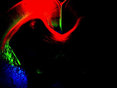
Studies reported that the corpus callosum, a large fiber tract that connects neurons in the right and left cerebral hemispheres, is diminished in Autism Spectrum Condition (ASC), and in some cases the corpus callosum does not even form. Understanding why these connections do not form properly in ASC is fundamental. Much of this wiring takes place during embryonic development. My laboratory is investigating the axonal deficits using ASC mouse animal models as well as brain slice culture

Figure 1: Corpus callosum projections labeled using DiI tracer [red] at embryonic day 17.
Neurogenesis and neuronal migration differences in ASC
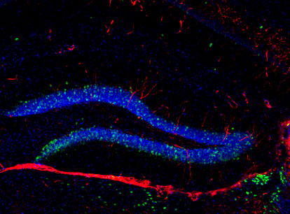
In ASC it has been previously reported that during brain development cells do not migrate to their final destination. In order to better understand what happens in ASC and what pathways are affected, we need to look at both neurogenesis and cell migration during brain development in ASC mouse models. We are particularly interested in investigating both neurogenesis and GABAergic interneuron migration during early brain development. Analyzing what pathways are affected during brain development in ASC and understanding when differences occur will help us elucidate how neurogenesis and cell migration are affected in ASC and what can be done to alleviate these differences.

Figure 2: Neurogenesis labeling in mouse hippocampal slice.
Lab Team
Céline Plachez, Ph.D.
Investigator
Karlie Menzel
Research Technician
Contact Information
Céline Plachez, Ph.D.
Investigator
Laboratory of Developmental Neuroscience
Program in Neuroscience
Hussman Institute for Autism
801 W. Baltimore Street / Suite 301
Baltimore MD 21201
443-860-2580 ext. 738
cplachez@hussmanautism.org
